What is cholesteatoma?
Cholesteatoma is a collection of a non-cancerous skin growth that develops within your middle ear. The middle ear is located just behind your ear drum and is responsible for the amplification of sound by the use of 3 small bones called ossicles. The most common cause of cholesteatoma is repeated middle ear infections. However, children can also suffer from cholesteatoma as a result of a birth defect. This condition usually develops as a sac or cyst that shed old skin which accumulates to increase the size of the cholesteatoma. In addition, it can destroy the delicate adjacent structures within the temporal bone- a pair of bones located at the lower outer portion of the skull which encloses the middle ear.
Cholesteatoma is one of the most common reason for ear surgery. In addition, it remains a relatively frequent cause of permanent or moderate conductive hearing loss. Conductive hearing loss occurs when there the passage of sound is blocked either in the ear canal or the middle ear. Generally, surgical removal of the growth is successful. However, it will continue to grow if not removed and may reappear even after surgical removal.
What are the causes of cholesteatoma?
There are 3 main types of cholesteatoma including:
- Congenital cholesteatoma: This is when a child has cholesteatoma as a result of a birth defect due to skin cells being trapped within the temporal bone while still in his/her mother’s womb. This develops into a cyst which contains fluids and dead skin cells. Conductive hearing loss is caused by obstruction of the eustachian tube which will lead to the accumulation of fluid within the middle ear. The eustachian tube is a tube which connects your middle ear to the back of your nose in order to equalise ear pressure. This tube also allows the removal of fluids or debris from the middle ear by use of gravity and some specialised cells called ciliated columnar epithelial cells. Furthermore, congenital cholesteatoma may expand posteriorly to encase the ossicles leading to conductive hearing loss.
- Primary acquired cholesteatoma: Eustachian tube dysfunction may create a vacuum within the middle ear resulting in retraction of the ear drum. This may lead bone erosion within the middle ear and destruction of the ossicles. This type of cholesteatoma can even damage your facial nerves. When the ear drum retracts, it may cause the formation of a cyst which may develop into a cholesteatoma. The growth will continue to enlarge as dead skin cells and fluids continue to accumulate.
- Secondary acquired cholesteatoma: This type of cholesteatoma occurs when your ear drum is injured due to frequent middle ear infections, trauma or surgical intervention.

What are the signs and symptoms of cholesteatoma?
The signs and symptoms of cholesteatoma include:
- Painless ear discharge: The hallmark of cholesteatoma is a painless, foul-smelling discharge from the ear. Infected cholesteatoma is very difficult to treat due to a lack of blood supply to the cholesteatoma as antibiotics cannot reach it properly. Antibiotic drop may be useful in this case. However, if the cholesteatoma is large and infected, it may recur despite aggressive treatment with antibiotics.
- Hearing loss: Another common symptoms of cholesteatoma is hearing loss. This occurs due to the accumulation of dead skin cells and fluids within the middle ear. When the ossicles are destroyed, this makes the hearing loss even worse.
- Vertigo: Advanced stages of cholesteatoma may produce a progressive symptoms called vertigo- this is when you feel that the surroundings are spinning around you which affects your balance. It may occur when the ossicles are damaged or when your inner ear is damaged- the inner ear is composed of a bony labyrinth which are responsible for balance and detection of sound.
- Facial nerve palsy: Cholesteatoma can sometime cause facial nerve paralysis.
- Perforated ear drum

There are some diseases which may resemble cholesteatoma and these include:
- Acute otitis media
- Chronic suppurative otitis media
- Otitis media with effusion
- Malignant otitis externa

Making a diagnosis
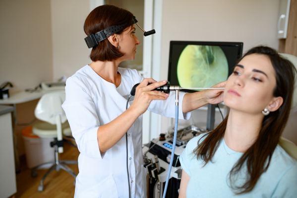
To make a diagnosis, your doctor will first take a detailed history from you to know more about your symptoms. After the history taking, your doctor will perform a thorough physical examination to look for signs of cholesteatoma. The physical examination involves your doctor using an otoscope to have direct visualisation of your ear drum and sometime inside your middle ear, if your ear drum is perforated. Your doctor will perform other tests in order to confirm the diagnosis and to determine the severity if the condition and these include:
- Audiometry: Audiometry is a test to determine your hearing ability. However, if the condition requires urgent surgery, this test can be done at a later stage.
- Computed Tomography (CT) scan: CT scan is the imaging of choice in the diagnosis of cholesteatoma as it can detect any damage caused to the temporal bone. However, even high-resolution CT scanning cannot determine the full extent of the condition. CT scans are especially useful in cases where the diagnosis is unclear, the patient does on want to undergo a surgical intervention or to assess for the presence of complications.
- Magnetic Resonance Imaging (MRI) scan: MRI scan is not used routinely and performed only in specific cases where there is presence of an abscess, inflammation of the tissue surrounding the brain (meningitis), inflammation of the facial nerve or signs of brain involvement. As this test does not use ionising radiation to produce images, it is useful in cases where the latter is contraindicated.


What are the treatments of cholesteatoma?
The treatment of cholesteatoma include:
- Surgical intervention: The only way to treat cholesteatoma is by removing it through a surgical intervention to prevent it from getting bigger and causing complications. After the cholesteatoma is removed, another surgical intervention might be necessary after some time to reconstruct the any damaged portions of the middle or inner ear.
- Antibiotics: Once cholesteatoma is diagnosed, antibiotic drops will be prescribed by your doctor to treat the infected cyst and reduce inflammation within the middle ear. This is usually done before surgery so that your surgeon is able to prepare for surgery better.

What are the complications of cholesteatoma?
If cholesteatoma is left untreated, the following complications may ensue:
- Mastoiditis: This is when the mastoid cells are infected. These cells are also known as air cells and are air-filled cavities within the mastoid process of the temporal bone of the skull. The signs and symptoms include fever, pain behind the ear, redness behind the ear and swelling of the earlobe (pinna).
- Facial nerve palsy: This complication may arise as a complication of the cholesteatoma or surgery. This affects the muscles involved in closing the eyes, smiling and raising the eyebrows. It can be partial or complete and immediately after surgery or after some days or weeks.
- Intracranial complications: Cholesteatoma can extend to the tissues surrounding the brain causing meningitis or cause intracranial abscesses which are life-threatening, requiring immediate intervention.
- Complete hearing loss in the affected ear after surgery.
- Difficulty to keep your balance.
- Persistent ear discharge after surgery which can be treated using a skin graft.

Expectations (prognosis)
Surgical intervention to treat cholesteatoma is usually successful. Unfortunately, the ossicles are sometimes so damaged that it cannot be completely restored back to normal. Nowadays, death from cholesteatoma is uncommon due to early detection, timely surgical intervention and appropriate antibiotic therapy.
Source:
J. Alastair, I. and Simon, M., 2016. Davidson's Essentials of Medicine. 2nd ed. London: ELSEVIER.
Parveen, K. and Michael, C., 2017. Kumar & Clarks Clinical Medicine. 9th ed. The Netherlands: ELSEVIER.
Patel, V., 2021. Cholesteatoma: Practice Essentials, Background, Etiology And Pathophysiology.
Isaacson, G., 2021. Cholesteatoma.


