What is pheochromocytoma?
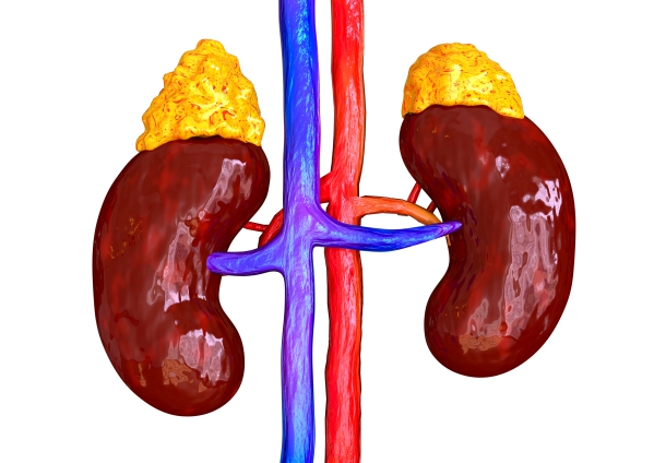
Pheochromocytoma is a tumour that develops in an adrenal glands. This tumour is usually benign (noncancerous). You have two adrenal glands, one sitting on top of each kidney. The tumour causes excessive release of the hormone called adrenaline which causes signs and symptoms such as increased heart rate, severe hypertension, sweating and headaches amongst others.
Around 10% of pheochromocytoma are discovered incidentally. This condition is rare, occurring in 0.05-0.2% of people suffering from hypertension. Pheochromocytomas can affect people of all races, however, black people are diagnosed less frequently. People aged between 30-50 years are more commonly affected by the disease.
What are causes and risk factors for pheochromocytoma?
Adrenaline is a hormone produced by the adrenal glands which prepares your body for a fight or flight response by increasing your heart rate and blood pressure. In pheochromocytoma, adrenaline is released in excess even when you are not in a threatened situation. There are several factors which can increase your risk of developing pheochromocytoma and these include:
- Multiple endocrine neoplasia type 2(MEN 2): MEN 2 is a genetic disorder associated with a high lifetime risk of developing tumours in various hormone-producing parts of the body.
- Von Hippel-Lindau disease: This condition results in tumours in various parts of the body including your pancreas, endocrine system, kidneys and central nervous system.
- Neurofibromatosis 1 (NF1): NF1 is a disease which results in tumours in your skin and optic nerve.
- Age: Pheochromocytoma can occur at any age. However, it is more common in people aged between 30 and 50 years.
- Race: This condition can affect people of all races. However, African Americans are less commonly affected.

What are the signs and symptoms of pheochromocytoma?
The release of excess adrenaline into your bloodstream from the tumour results in the following signs and symptoms:
- Palpitations: A feeling of having a fast-beating heart or pounding heart.
- Heavy sweating (diaphoresis)
- Headache
- Tremor: An involuntary, rhythmic muscle contraction that results in shaking movements in one or multiple body parts.
- Nausea
- Tiredness
- Pallor of the face
- High blood pressure: The hypertension associated with pheochromocytoma is episodic and volatile
- Shortness of breath
- Anxiety: Having a feeling of hopelessness or sense of doom during the pheochromocytoma attack.
- Pain in the upper abdomen
- Flank pain
- Weight loss
- Constipation
- Increased heart rate
People suffering from pheochromocytoma often complain of spells characterized by headaches, palpitations, diaphoresis and hypertension. These spells vary and tends to get worse with time. Certain activities or conditions may worsen your symptoms and these include physical exertion, stress, changes in body position and surgery amongst others. In addition, some foods may make your symptoms worse and these include some cheeses, beers, wines and chocolate. This is because these foods contain tyramine which is often found in aged, pickled, fermented, cured or spoiled foods. Furthermore, certain medications can make your symptoms worse and these include monoamine oxidase inhibitors (MAOIs) and stimulants such as amphetamine and cocaine.
Making a diagnosis
To make a diagnosis, your doctor will first take a detailed history from you to know more about your symptoms. After the history taking, your doctor will perform a thorough physical examination to look for signs of pheochromocytoma. He/she will order some tests in order to confirm the diagnosis and these include:
- 24 hour urine test: This test involves the collection of your urine over a 24 hour period to test for creatinine and adrenaline to determine whether there is excess adrenaline production.
- Blood tests: A blood sample may be taken to measure the level of adrenaline in your blood.
- Clonidine suppression test: This test can be used to diagnose pheochromocytoma. Clonidine is a drug that usually decreases the level of adrenaline. Clonidine is given orally and the level of adrenaline is measured. If the level of adrenaline is not suppressed, the diagnosis of pheochromocytoma can be made.
- Abdominal Computed Tomography (CT) scan: Abdominal CT scan is performed to detect any adrenal masses which can be used to make the diagnosis of pheochromocytoma.
- Magnetic Resonance Imaging (MRI) scan: MRI scan is referred in children and pregnant women as no ionising radiation is involved. In addition, this test is very accurate in detecting pheochromocytomas.
- Scintigraphy: This test is a nuclear medicine scan that uses iodine-123 metaiodobenzylguanidine (MIBG). The MIBG scan is used when the diagnosis cannot be made via CT or MRI scan despite abnormal levels of adrenaline.
- Positron emission tomography (PET) scan: PET scan can be used to detect pheochromocytomas.
- Genetic testing: This test can be used to rule out a familial syndrome in someone suffering from pheochromocytoma.
What are the treatments of pheochromocytoma?
The management of pheochromocytoma mainly involves surgery to remove the tumour. However, the preoperative preparations are very important to increase the rate of recovery. Alpha blockers, beta blockers and a salt restriction diet are important to control the hypertension associated with pheochromocytoma. Alpha blockers work by relaxing your blood vessels in an attempt to lower your blood pressure. Beta blockers work by making your heart beat slower and with less force.
Surgical removal of the tumour results in cure of the hypertension. Laparoscopic adrenalectomy is the surgical procedure of choice when the tumour is less than 8cm. This procedure involves your surgeon making 3 small incisions in your abdomen in order to pass tools and a tube containing a camera at its end.
If the tumour is malignant (cancerous), MIBG, chemotherapy and radiotherapy can be used to kill the cancer cells. However, malignant pheochromocytomas are very rare.
What are the complications of pheochromocytoma?
The complications associated with pheochromocytoma occurs mainly due to severe and longstanding hypertension. If left untreated, the following complications may ensue:
- Stroke: This condition occurs when a blood vessel in your brain breaks due to hypertension which may be life-threatening.
- Cardiomyopathy: This is a condition affecting your heart muscles which makes it harder for your heart to pump blood to other parts of the body.
- Kidney failure
- Eye problems: Hypertension may cause the small blood vessels within your eye to break leading to haemorrhages.
- Myocarditis: This is when there is inflammation of your heart muscle.
- Pulmonary edema: Pulmonary edema is the accumulation of fluids in your lungs.
Prognosis
The 5-year survival rate for people suffering from noncancerous pheochromocytoma is greater than 95%. Unfortunately, people suffering from cancerous pheochromocytoma have a 5-year survival rate of only 50%. Pheochromocytoma is a rare but deadly condition which is why it is of utmost importance to seek medical advice if you have symptoms of this disease.
Source:
J. Alastair, I. and Simon, M., 2016. Davidson's Essentials of Medicine. 2nd ed. London: ELSEVIER.
Parveen, K. and Michael, C., 2017. Kumar & Clarks Clinical Medicine. 9th ed. The Netherlands: ELSEVIER
Blake, M., 2020. Pheochromocytoma Clinical Presentation.
Blake, M., 2020. Pheochromocytoma Workup.
Blake, M., 2020. Pheochromocytoma Treatment & Management.