What is Uveitis?
The uveal tract is a structure of the eye. It is a layer which is found just beneath the white part of the eye known as the sclera. When this layer is inflamed, the condition is known as uveitis. The uveal tract is divided into the anterior (found in the front) and the posterior (found at the back) compartment. The anterior compartment is composed of the iris and the ciliary body. The iris is the coloured part of your eye that you can see when you look at it in the mirror. The ciliary body is the part of the eye which is responsible to change the size of the pupil, the shape of the lens as well as to produce the fluid that fills the eyeball. The posterior compartment is composed of the choroid. It is a layer that provides nutrients to surrounding layers of the eye.
Most of the time, the exact cause of uveitis is unknown. However, in some cases, it may be triggered by trauma, immune, infectious or genetic mechanisms. Uveitis is a common cause of preventable blindness, especially when the posterior part is involved. When the anterior compartment is affected, it is the most common form to present at the emergency department. In the United States, around 25-52 people per 100,000 are affected by the condition per year. Worldwide, Finland is one of the places where there is the highest incidence of uveitis recorded. It is a condition that occurs equally in males and females. However, those aged between 20-50 years are most commonly affected. The prognosis of uveitis is usually excellent with prompt and appropriate treatment.

What are the causes of uveitis?
The exact cause of uveitis is unknown. However, there are several factors that may contribute in the development of the disease. These include genetic, traumatic or infectious factors that can promote or trigger the inflammation of the uveal tract. Uveitis is also commonly associated with other medical conditions including inflammatory bowel disease, rheumatoid arthritis, systemic lupus erythematosus, sarcoidosis, tuberculosis, syphilis and acquired immunodeficiency syndrome (AIDS).
When trauma occurs to the eye, pathogens are more likely to get into the eye at the site of injury. This can trigger an inflammatory response in the eye leading to uveitis. Other infections that may result in uveitis (especially anterior compartment) is Herpes virus infection.
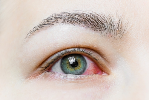
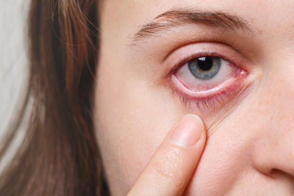
What are the signs and symptoms of Uveitis?
There are many signs and symptoms that can arise from uveitis. These include:
- Pain
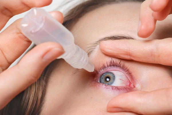
- Red eye
- Photophobia: Increased and excessive sensitivity to light
- Excessive tearing
- Decreased vision
- Blurry vision
- Floaters: Small dark shapes that float across your vision
It is important to seek medical help as soon as possible if you experience the above symptoms as there are many eye conditions that can present with same.
What are the conditions that can mimic uveitis?
The following conditions can present with the same symptoms as uveitis:
- Glaucoma: When the pressure inside the eye increases due to inadequate drainage of fluid in the ocular space
- Acute conjunctivitis
- Ulceration of the cornea
- Presence of a foreign body in the eye
- Inflammation of the sclera (white part of the eye)
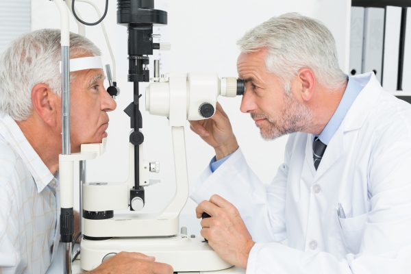
How is the diagnosis of uveitis made?
To make the diagnosis of uveitis, your doctor will begin by asking you a series of questions to know more about your symptoms. He/she will then perform a physical examination focusing on the eye. The eye examination includes the following:
- Vision assessment: Your doctor will check the response of your pupils to light. He/she will also assess your visual acuity which is how well you can see/read.
- Tonometry: In this procedure, a special device is used to measure the pressure inside your eye (intraocular pressure). Prior to the procedure, eyedrops are used to numb your eyes so that the procedure goes painless.
- Slit lamp examination: In this procedure, a special apparatus, involving magnifying lenses and light sources, is used to examine the eye.
- Ophthalmoscopy: This is also known as Fundoscopy. To carry out this examination, your doctor will first apply eyedrops to your eyes that will dilate your pupils to enable proper examination of the back of the eye (fundus). An ophthalmoscope is used to perform the examination.
- Other imaging tests: In some cases, your doctor may request some other tests including optical coherence tomography (OCT), fluorescein angiography, analysis of the fluid in the eyeball or computed tomography scans.
How is uveitis treated?
If you present with uveitis in the emergency department, you will be referred to a specialist (ophthalmologist), after having been provided with pain killers.
The main goals of treatment are pain and inflammation reduction with cycloplegics and corticosteroids:
- Cycloplegics: These medications are used to dilate the pupil as well as controlling spasms of the iris and ciliary body. This contributes in alleviating the pain associated with uveitis. These are available as eyedrops. Some examples include cyclopentolate, homatropine, scopolamine and tropicamide.
- Topical corticosteroids: These drugs work by alleviating inflammation associated with uveitis. These are also used in the form of eyedrops. However, if the posterior compartment is involved, injections of corticosteroids in the eye may be necessary. In some cases, corticosteroids may be taken by mouth. An example include prednisolone. However, some side effects include: increased intraocular pressure, formation of cataract or steroid induced glaucoma.
- Antibiotics or antivirals: If the uveitis is found to be due to a bacterial or viral infection, antibiotics or antivirals may be prescribed respectively.
Surgery is not usually warranted in the treatment of uveitis.
What are the complications of uveitis?
The inflammation or use of medication (topical corticosteroid) can result in an increased pressure inside the eyeball. This is the most important complication of uveitis. If left untreated, the increased intraocular pressure can result in damage of the optic nerve leading to permanent vision loss.
Source:
Muchatuta, M., 2019. Iritis and Uveitis.
Dunn JP. Uveitis. Prim Care. 2015 Sep. 42 (3):305-23.
Wills Eye Hospital. The Wills Eye Manual: Office and Emergency Room Diagnosis and Treatment of Eye Disease. 5th ed. Philadelphia, Pa: Lippincott; 2008.
Islam N, Pavesio C. Uveitis (acute anterior). Clin Evid (Online). November 2009. 04:705:
Yanoff and Duker. Uveitis and other intraocular inflammations. Ophthalmology. 3rd ed. Mosby; 2008.