What is nephrolithiasis?
Nephrolithiasis, also known as kidney or ureteric stone, refers to when a calculi or stone is formed in the urinary tract. It most commonly originates from the kidney and gets lodged in the ureters which are tube like structures carrying urine from the kidneys to the bladder. Most of the stones contain calcium.
This condition can be very painful as the stone stretches the ureters and causes obstruction to urine flow. It is a common disease which affects around 1 in 11 people in the United States. Its incidence and prevalence have increased during the last years. This condition occurs all around the world. It most commonly affects people aged between 20-49 years but can affect anyone at any age. Besides, nephrolithiasis has been found to be more common in men
The initial management of kidney stones starts with proper diagnosis and rapid initiation of treatment. The treatment of nephrolithiasis depends on the type and size of stone present in the urinary tract.
Causes of nephrolithiasis
There are several factors known to be causing the formation of kidney stones. These include:
- Low fluid intake: When you do not consume enough water, your urine tends to become more concentrated. Hence, it is more likely to form stones.
- High amount of calcium in urine: If you consume too much calcium or your body is absorbing excessive amount of calcium, you end up with urine high in calcium. High amount of calcium in the urine favours the formation of stones.

There are different types of stones known depending on their composition, namely:
- Calcium stones: these are the most common types of stones
- Struvite stones also known as infection stones
- Uric acid stones: these stones are common in people who consume a lot of proteins
- Cysteine stones: this is common in people who have a hereditary problem which causes the kidneys to excrete too much of a protein called cysteine.
Risk factors
The following factors put you more at risk of having nephrolithiasis:
- Diet rich in protein: You are at increased risk of ending up with uric acid stones if you consume at protein rich diet.
- Dehydration: Concentrated urine can precipitate the formation of kidney stones
- Being obese: Obesity increases the likelihood of getting
- Living in warm and dry areas
- A personal or family history of kidney stones: If you had kidney stones in the past or have close family members who do, you are more likely to have kidney stones.
- Recurrent urinary tract infections: Recurrent infections can predispose you to struvite stones.
Signs and symptoms of Nephrolithiasis
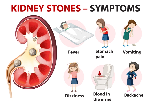
Whenever a large stone is lodged within your urinary tract, it causes stretching of the tubes and obstruction which can lead to several signs and symptoms such as:

- Severe and sharp pain on the flank which radiates to the groin area
- Pain coming in attacks
- Blood in urine
- Painful urination
- Vomiting
- Nausea
- Foul smelling urine in cases of urinary tract infections
- Fever if coexisting infection
When your doctor will examine you, he/she may find that you are not able to stay still and your abdomen is tender on palpation. Your kidney may also be enlarged due to obstruction to urine flow.
Smaller stones on the other hand may pass spontaneously on passing urine.

Making a diagnosis
With a proper history, your doctor may already suspect kidney stones. However, certain tests will have to be done to confirm the diagnosis or rule out other conditions that can present in the same way. These tests include:
- Blood tests: In blood tests it can be found whether there is an excessive amount of calcium or uric acid in the body which can promote the formation of stones. Functioning of the kidney can also be assessed through blood tests.
- Urine tests: A sample of your urine will be taken for analysis to look for the presence of stone-forming minerals or infection. Presence of blood in the urine can also help in the diagnosis.
- Abdominal X-ray: Stones which contain calcium can be visible on abdominal X-ray. It is a quick and inexpensive way to identify the presence of nephrolithiasis. However, other types of stones may not be visible on plain abdominal X-ray.
- Ultrasound: This is mostly used in pregnant women or may be used in combination with abdominal X-ray in other individuals. It also allows identification of stones not visible on X-ray.
- Computerized tomography scan: This imaging modality can allow identification of even tiny stones which can be missed on X-ray or Ultrasound.
- Intravenous pyelography: This is the test of choice to make the diagnosis of nephrolithiasis. It allows a proper visualization of the whole urinary system and how the kidney is functioning.

Using imaging modalities, the exact location and size of the stone will be known and the treatment will depend on these factors.
Management of nephrolithiasis
Small kidney stones may pass spontaneously without any surgery. However, the following measures may help:
- Increasing your water intake: This can facilitate the passage of small stones as well as causing a dilution of urine, hence decreasing the risk of stone formation.
- Pain killers: Some degree of pain may be present when a small stone will pass through urine. To relieve the pain, some pain killers may be given to you.
- Alpha Blockers: these medications are given to relax the muscles in your ureter to allow easier passage of the stone. An example is tamsulosin.

In the case of larger stones, it is impossible that they pass on their own. Therefore, surgical procedures may be warranted:
- Extracorporeal shock wave lithotripsy: In this procedure, sound waves are used to cause vibration of the stones. When this happens, the stones may break into smaller pieces which can pass through urine.
- Percutaneous nephrolithotomy: In this procedure, a small incision is made on your back and instruments are inserted through it to reach the kidney. The kidney stones are then removed using these special instruments.
- Ureteroscope: A thin tube is inserted through your urethra up to your ureter. Once the level of the stone is reached, the stone is broken into pieces using special equipments through the tube. The pieces of stone will subsequently pass through urine.
- Placement of stent: In cases where there is the presence of an obstructing stone as well as infection, a stent is placed and treatment of the infection is initiated till it resolves. This provides temporary relief from the obstruction. Removal of the stone will be done after resolution of the infection.
Complications of nephrolithiasis
Several complications can arise from kidney stones including:
 Formation of an abscess
- Infections of the kidney that affects kidney function
- Fistula formation which is a connection between the urinary tract and surrounding structures
- Scarring and narrowing of the ureters
- Infection spreading in the blood
- Kidney failure if left untreated
Prevention
Some preventive measures include:
- Drinking about 2 litres of water per day
- Decreasing the amount of salt and animal protein in your diet
- Using calcium supplements with precaution but do not restrict your dietary calcium intake
- Thiazide diuretics may be used in people with high urine calcium levels and recurrent calcium stones
- Potassium citrate can be used in people with recurrent uric acid and cysteine stones
Prognosis
About 80-85% of stones will pass on their own while about 20% will need to be admitted due to severe pain and inability to pass the stone. According to studies made, increasing fluid intake and modifying your diet can decrease the recurrence rate by 60%.

Source:
Dave, C., 2020. Nephrolithiasis.
Parveen, K. and Michael, C., 2017. Kumar & Clark's Clinical Medicine. 9th ed. The Netherlands: ELSEVIER.


