
What is retinal detachment?
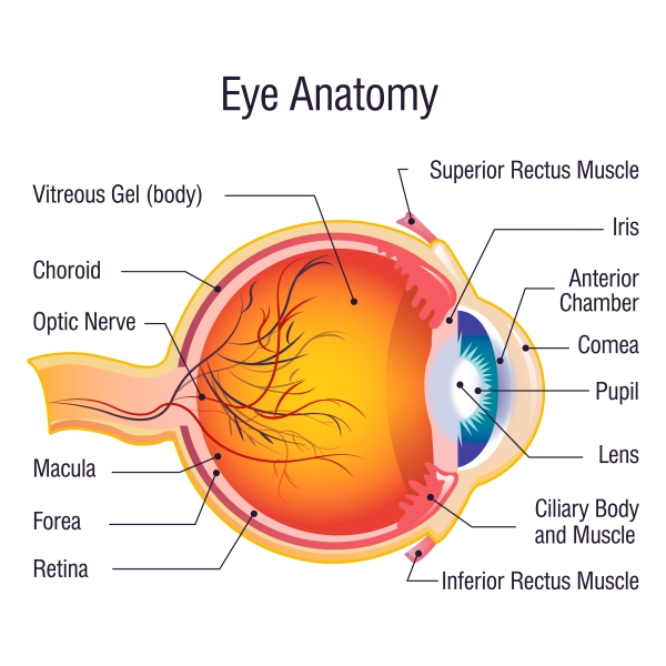
Retinal detachment is an ocular emergency whereby your retina at the back of your eye detaches from its normal position. The retina is a thin layer of tissue found at the back of your eye. Its function is to capture light from the lens and convert it into signals. The retina is then responsible to send these signals to the brain for interpretation and formation of images of what we can see. Therefore, the retina plays an essential role in vision.
When the retina is detached from its normal position, it is cut off from oxygen and essential nutrients for it to function properly. If retinal detachment is left untreated for a long period of time, permanent loss of vision may ensue. The occurrence of retinal detachment increases with increasing age. It usually occurs in people aged between 40 and 70 years. It also occurs equally in men and women.
What are the causes of retinal detachment?
There are several factors that can lead to retinal detachment. In some cases, a hole or tear may be present in the retina. This allows passage of fluid through the hole and accumulate under the retinal layer. This causes a pulling force on the retina, causing its detachment from the underlying layer. This is a common mechanism in old people. The detached retina loses its oxygen and nutrient supply, resulting in visual loss.

Another mechanism may be by the formation of scar tissue on the retinal layer, pulling the retina away from the back of the eye. This is known as tractional retinal detachment. This is a common cause of retinal detachment in people with uncontrolled diabetes.
Retinal detachment can also arise from an accumulation of fluid underneath the retinal layer in the absence of any hole or tear. This may be due to ocular injury, tumours and inflammation in the eye.
What are the risk factors for retinal detachment?
You are more at risk of having retinal detachment if you:
- Are aged above 40 years
- Have a history of retinal detachment
- Have close family members with retinal detachment
- Are myopic which means that you see objects that are near to you clearly while far away objects appear blurry
- Have a history of eye surgery
- Had sustained an eye injury
What are the signs and symptoms of retinal detachment?
Retinal detachment can present with a variety of signs and symptoms. Some of them may appear suddenly while others may take time to become apparent. These include:
- Sensation of a flashing light
- Seeing floating or dark spots in your field of vision (also known as floaters)
- Loss of vision which can be described as cloudy, irregular or curtain-like
- Blurred vision
In normal vision, you may sometimes notice some tiny floater. This is completely normal. However, if they appear suddenly, become more frequent and very large, this means that there is an eye problem. The floaters may have the appearance of a spider web, a single large spot or multiple small spots.
As soon as you notice having floaters, flashes of light or loss of vision, seek medical help immediately. If not treated as soon as possible, complete loss of vision may occur.
How is the diagnosis of retinal detachment made?
There are no laboratory tests that help in making the diagnosis of retinal detachment. These are done only if surgery will be required, in order to know about your general condition and whether you are fit for surgery.
Imaging tests are more useful to aid in the diagnosis of retinal detachment. These include:
- Eye ultrasound: This test can be done at bedside to see whether retinal detachment is present. However, some other conditions such as posterior vitreous detachment and vitreous haemorrhage may mimic the appearance of retinal detachment in ultrasound.
- Dilated eye exam: In this procedure, your doctor will apply eye drops to your eye which will help to keep your pupil dilated. This will allow your doctor to use some special devices to look into your eye and have a look at your retina. Retinal detachment may be identified on dilated eye exam.
How is retinal detachment treated?
Once the diagnosis of retinal detachment is made, treatment should be given as soon as possible to prevent further damage to the eye. The treatment choice will depend on the type of retinal detachment present. Your doctor knows best on what treatment modality to choose depending on the stage of the retinal detachment. Options include the following:
- Laser photocoagulation: In this procedure, a laser beam is projected into your eye to make burns around retinal tears present, thus attaching the retina firmly onto the underlying layers.
- Cryopexy: In this procedure, a freezing probe is applied over tears on the retinal surface in order to secure the retinal layer onto the back of the eye.
- Injecting a bubble of gas into the eye: In retinal detachment, this can be done as a means to push back the detached retinal layer into its original place. This is known as pneumatic retinopexy. This stops further accumulation of fluid underneath the retina, hence stopping the progression of the detachment. Fluid that is already collected under the retina gets absorbed on its own. With time, the bubble of gas will also go away without any further intervention.
- Scleral buckling: In this procedure, a surgeon sutures a piece of silicone to the white of your eye at the affected area. This decreases the tension on the retina.
- Vitrectomy: This is a procedure involving the removal of the gel-like liquid inside of your eye known as the vitreous, as the latter may be pulling on the retina worsening the detachment. The vitreous is then replaced by another substance such as air, gas or silicone oil. This will help in flattening the retina over the underlying structures. This procedure can even be combined with other surgical procedures.
What are the complications of retinal detachment?
The most common and serious complication of retinal detachment is loss of vision if left untreated. This can be avoided if you seek medical help as soon as possible after the appearance of symptoms.
What is the prognosis for retinal detachment?
Around 90% of treatments for retinal detachment are successful. However, some people may require further treatment. In some cases, the retina cannot be attached and hence this leads to progressive loss of vision. Since the retina is a tissue containing important neurone networks, the prognosis can be difficult to predict.
Source:
Pandya, H., 2018. Retinal Detachment: Practice Essentials, Background, Pathophysiology.
Arroyo, J., 2020. Retinal Detachment.