What is Tinea capitis?
Tinea Capitis is a superficial fungal infection of the skin of the scalp, eyebrows and eyelashes. It characteristically attack hair shafts and follicles. This condition is also known as ringworm of the scalp. It belongs to the same family as tinea corporis (ringworm of the glabrous skin), tinea cruris (ringworm of the groin), tinea unguium (ringworm of the nails), tinea pedis (ringworm of the feet) and tinea manuum (ringworm of the hand).
With time, the incidence of the disease has been increasing around the world. It is mostly a disease that affects preadolescents. It typically arises among those aged between 5 to 10 years. Tinea capitis is responsible for around 92.5% of fungal skin infections in children aged less than 10 years. It occurs less occasionally among adults but can sometimes arise in elderly people. Tinea capitis is also widespread in some urban areas in North America, Central America and South America. It is also common in some parts of Africa and India. Tinea capitis is also more common among males compared to females. However, it depends on the exact pathogen causing the disease which may be more common in females compared to males.
What are the causes of Tinea capitis?
Tinea capitis is caused by a fungus. The most common fungi responsible for the development of tinea capitis in the United States is T tonsurans. It is also the most common causative pathogen in Canada, Mexico and Central America. The fungus can also be found in animals. Transmission from animals to humans occurs following direct contact with infected animals. Human to human transmission is the main mode of transmission of the disease. One person can transmit the infection to another through direct skin-to-skin contact. It can also spread through fomites, which are contaminated surfaces, clothing, towels, bed sheets, combs or hair brushes.

Once the fungi gets into the scalp, it invades the superficial layer of skin and attacks the hair shaft. The infected hair becomes brittle and breaks off easily, leaving behind patches of missing hair.
What are the risk factors for Tinea capitis?
The following factors increases the risk of having tinea capitis:
- Being a toddler or a school-aged child.
- Being exposed to infected people, especially having close skin-to-skin contact.
- Being exposed to infected animals such as cats or dogs. Transmission can occur during petting or touching the animal.
What are the signs and symptoms of Tinea capitis?
The following signs and symptoms may be present in tinea capitis:
- One or more ring-shaped patches of skin covered with scales which slowly enlarge and expand
- Ring-shaped lesions that coalesce with each other
- Lesion which starts as a small reddish bump around the hair shaft on the scalp
- Brittle and discoloured hair which breaks off a few millimetres above the scalp surface
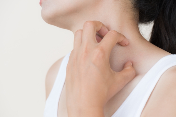
- Itching of the lesions
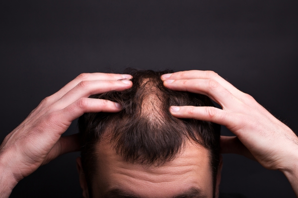
- Hair loss in areas affected (alopecia)
- Deep boggy red areas (if an abscess has developed) also known as a kerion
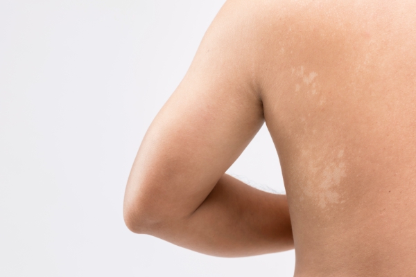
Tinea capitis usually affects the scalp, eyebrows and eyelashes.

How is the diagnosis of Tinea capitis made?
To make the diagnosis of Tinea capitis, your doctor will first start by asking you a series of questions to know more about your symptoms. He/she will then proceed with a physical examination, focusing on the affected area of skin. Most of the time, the history taking and physical examination is sufficient to make the diagnosis. However, in some cases, your doctor may request some special tests to confirm the diagnosis. A sample of affected skin or hair may be taken for further evaluation under the microscope. The fungus may be identified using these special tests. Examination using KOH (potassium hydroxide) solution can also be useful for the proper diagnosis.
How is Tinea capitis treated?
The choice of treatment for tinea capitis depends on the exact causative pathogen, the degree of involvement and the general health of the individual affected. As soon as the diagnosis has been made, treatment should be initiated. Treatment options include the following:
- Antifungal medications: In some cases, your doctor may prescribe oral antifungals such as griseofulvin. The recommended dose is based on body weight. The treatment may last for about 4 to 6 weeks. Some newer alternatives include itraconazole and terbinafine.
- Selenium sulfide shampoo: Your doctor may recommend the use of selenium sulfide shampoos to reduce the risk of spreading the infection to other people.
How can Tinea capitis be prevented?
Siblings and playmates of those who are known to be infected should avoid close contact or sharing of toys or other objects. People who are infected should have their pets inspected and treated accordingly.
What are the possible complications of Tinea capitis?
The main complication of tinea capitis is breaking off of hair as well as scaring alopecia. This is when the lesion heals but hair no longer grows on that region due to the scarring. In some cases, abscess can also develop with time. Tinea capitis mostly causes psychological distress to the affected individual. Since it mainly occurs among school aged children, bullying is a common issue.
What is the prognosis for Tinea capitis?
With appropriate treatment, tinea capitis usually resolve completely carrying an excellent prognosis. People who have not received adequate treatment or who have tinea capitis that is resistant to treatment are at increased risk of having an abscess development. This is known as a kerion. Despite having ongoing treatment, fungal spores are continuously shed for several months. Some factor may contribute to treatment failure including reinfection, pathogen being insensitive to treatment, medication not being absorbed optimally or the affected person is not complying with the treatment as advised.

Source:
Handler, M., 2020. Tinea Capitis
Rayala BZ, Morrell DS. Common Skin Conditions in Children: Skin Infections. FP Essent. 2017 Feb. 453:26-32.
Seebacher C, Bouchara JP, Mignon B. Updates on the epidemiology of dermatophyte infections. Mycopathologia. 2008 Nov-Dec. 166(5-6):335-52.
Thakur R. Tinea capitis in Botswana. Clin Cosmet Investig Dermatol. 2013. 6:37-41.
Fuller LC, Child FJ, Midgley G, Higgins EM. Diagnosis and management of scalp ringworm. BMJ. 2003 Mar 8. 326 (7388):539-41.